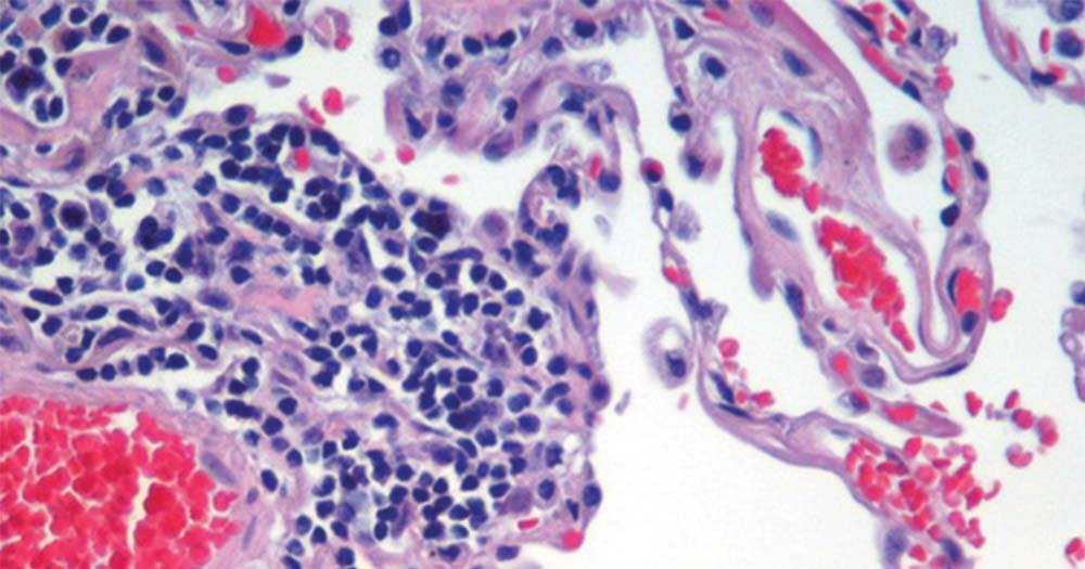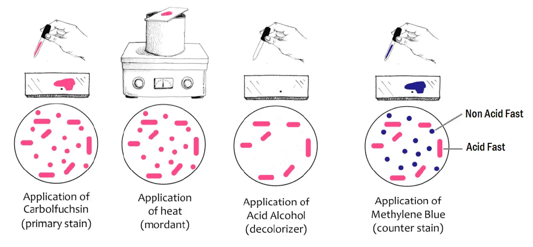

Keywords: detection eosinophil histochemistry lung mouse RSV. This is a quick method for the identification of Cryptococcus neoformans. This forms a zone of clearance known as a halo, around the cells. Outcome was poor (mean survival time, 23 weeks) despite treatment. Formultiple parameters such as eosinophil detection, specificity, and contrast with background tissues, the Sirius Red followed by Congo Red andModified Hematoxylin and Eosin methods were useful, each with their own staining qualities. Using India Ink for visualizing the cerebral spinal fluid, indicating that particles of ink pigments can not enter the cell capsule surrounding the yeast cells.

Diffuse interstitial opacities (70.5%), focal interstitial abnormalities, alveolar opacities, adenopathies, cavitary lesions, and pleural effusions were evident. Indian ink preparation of both samples showed plenty of capsulated budding yeast cells morphologically similar to Cryptococcus Figure - 2. Clinical manifestations of pulmonary cryptococcosis included fever (94%), cough (71%), dyspnea (7%), expectoration (4%), chest pain (2%), and hemoptysis (1%). The large particles of ink will not penetrate the tight layers of the capsule or stain the bacterium. One very simple approach is mixing cells in a preparation of India ink. The CD4⁺ lymphocyte count was low in all cases (median, 24/mm³). History Since the 1900s various methods have been devised to observe bacterial capsules (7). All but one of the 27 patients had detectable CA in serum. This capsule is comprised primarily of glucuronoxylomannan, which is a polysaccharide of. neoformans was cultured from bronchoalveolar lavage (BAL) or pleural fluid in 25 cases the remaining two patients had cryptococcal antigen (CA) detected in BAL fluid and C. The India ink does not penetrate the cryptococcal carbohydrate capsule. The latex agglutination test is a more sensitive method but may still yield. Twenty-seven patients (32%) had pulmonary cryptococcosis. Sputum for identification of infectious pulmonary tuberculosis (limited sensitivity). Although negative staining like India ink and nigrosin are most widely used. We reviewed the records of 85 patients infected with both human immunodeficiency virus and Cryptococcus neoformans.


 0 kommentar(er)
